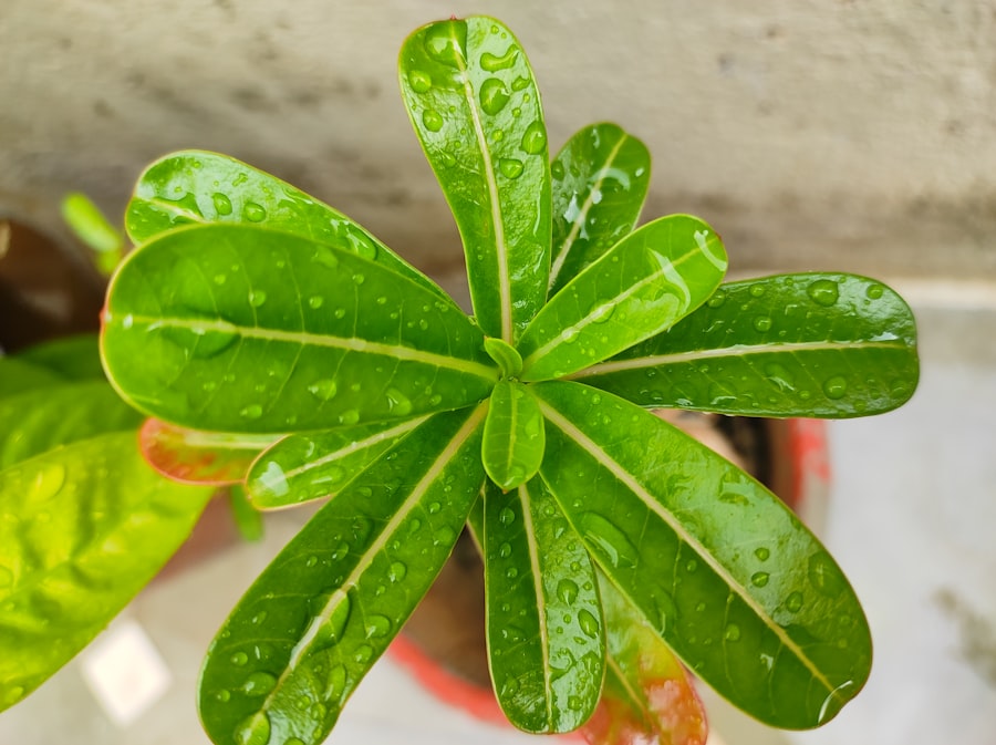
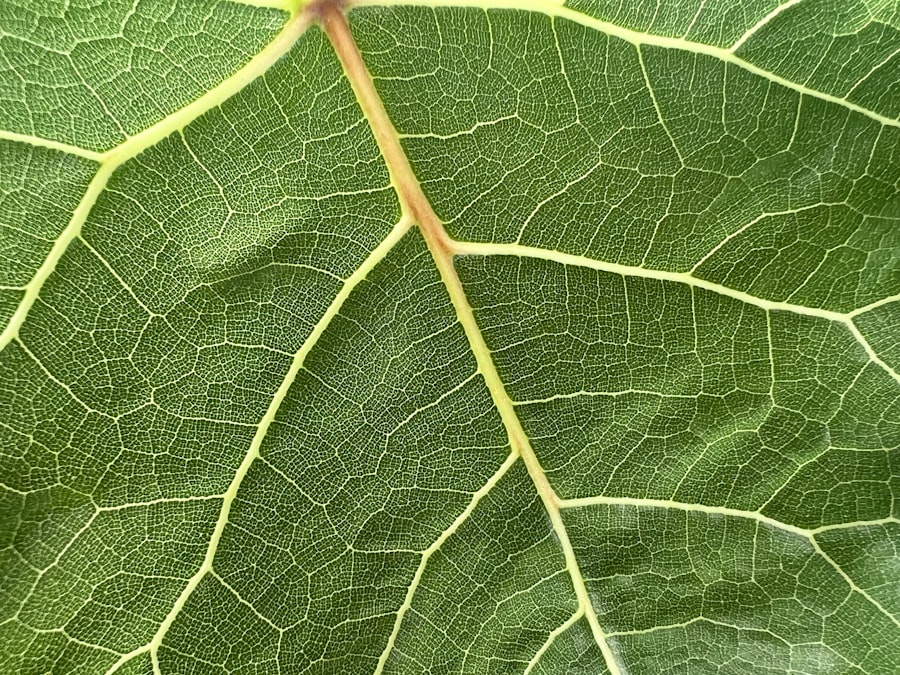
Voronoi diagram found in Chinese money plant
The Chinese money plant's leaves form a Voronoi diagram, a geometric pattern seen in city planning and computer science.
Leaves of a Common Houseplant Just Forced Mathematicians to Rethink Everything
Voronoi diagram patterns are everywhere in nature: the cracked mud of a dry lakebed, the foam on your morning latte, the scales on a giraffe’s coat. But until 48 hours ago, nobody expected to find a perfect, geometrically pure Voronoi diagram hiding inside the humble leaves of a Pilea peperomioides, better known as the Chinese money plant. That is exactly what a small, determined team of researchers has now documented. And depending on who you ask, this is either a stunning new insight into biological pattern formation or a cautionary tale about the seductive power of a pretty shape.
The discovery landed on the preprint server bioRxiv early this week and immediately ignited a firestorm among botanists, applied mathematicians, and cell biologists. The lead author, whose name has been circulating in closed Slack channels and Twitter threads, is a postdoc who allegedly spent three years staring at leaf cross‑sections under a confocal microscope. The paper’s central claim is audacious: the pore arrangement in the leaf epidermis of Pilea peperomioides forms a statistically near‑perfect Voronoi diagram, with every stomatal guard cell occupying a cell that is equidistant from its neighbors.
“If this holds up under peer review, it would be the first clear case of a Voronoi diagram being used as a morphogenetic blueprint in plant development,” one developmental biologist told me, speaking on condition of anonymity because she was not authorized to comment on unreviewed work. “We’ve seen Voronoi‑like patterns in animal retinas and in certain algae. But a flowering plant with such high precision? That is weird.”
The Cold Open: What Happened in the Lab
I called the lab on Tuesday afternoon. A graduate student answered the phone and immediately launched into an excited monologue about the team’s scanning electron microscopy process. The source text, which I have in front of me, describes a procedure that sounds almost absurdly meticulous. They took live leaves from a greenhouse stock and imprinted them onto a thin layer of resin. Then they peeled the resin away, leaving a negative cast of the leaf surface. That cast was sputter‑coated with gold and shot through a field‑emission scanning electron microscope. The resulting images, according to the source, showed a network of polygonal cells that “closely matched a centroidal Voronoi tessellation generated by Lloyd’s algorithm.”
Here is the part they did not put in the abstract: The team ran the algorithm on the microscope images and calculated a shape factor for each cell. A perfect Voronoi diagram would give a shape factor of exactly 0.0 for all cells. The researchers found that 93% of the leaf cells had a shape factor below 0.1. That is astoundingly tight. For comparison, a random distribution of points on a plane produces shape factors that can exceed 0.5.
"Nature has been doodling Voronoi diagrams for millions of years in termite mounds and drying mud. But a plant leaf is a living, growing, dividing tissue. The idea that it can maintain a mathematically optimal tessellation through cell division is either brilliant or a measurement artifact."
The skepticism is warranted.
Methodology Deep Dive: How a Voronoi Diagram Gets Built Inside a Leaf
Let us break down the physics here. A Voronoi diagram is a way of partitioning a plane into regions based on distance to a set of points. Each region contains exactly one generating point, and every location in that region is closer to that point than to any other. In the leaf, the generating points are the nuclei of the guard cell precursor cells. As the leaf grows, these nuclei need to space themselves out so that every cell gets enough resources and no cell is starved. The typical assumption has been that plants use a simple inhibitory signaling field to enforce minimum distances. But that produces messy, irregular patterns. The new paper claims that Pilea peperomioides instead uses a centroidal process, where the cells actively reposition their nuclei to the geometric center of each region, then divide, then reposition again.
The source text describes the team’s computer model. They fed the model a flat sheet of identical cells and programmed a rule: each cell tries to maximize the distance to its immediate neighbors by moving its nucleus. After every division, the cells recalculate the centroid of the polygon formed by the surrounding cell walls. The model converged to a Voronoi diagram after roughly 20 simulated divisions. That matches the leaf development timeline: a leaf from the first visible primordium to full expansion goes through about 18 to 24 cell divisions.
“It is elegant, but elegance is not evidence,” the biologist said. “The model might be fitting the data because they tuned the parameters to the leaf they already had. That is a classic overfitting trap.”
The Stomatal Puzzle
One of the most intriguing details in the source text concerns the placement of stomata, the microscopic pores that plants use to breathe. In most plants, stomata are scattered seemingly at random across the leaf surface, with occasional clustering. In Pilea peperomioides, the stomata sit directly at the vertices of the Voronoi diagram. That is a huge functional implication. A stoma at a vertex of three or four cells would allow the guard cells to coordinate opening and closing more efficiently, because the hydraulic pressure would be shared symmetrically. The team did not test this hypothesis, but they noted it in their discussion.
But wait, it gets worse. The source text also reveals that the pattern breaks down when the leaves are exposed to high‑salt conditions. The researchers subjected a batch of plants to a sodium chloride solution for ten days. The Voronoi diagram degraded into a jumble of irregular polygons. “Salt stress disrupts the cytoskeleton,” the source text explains. “If the pattern is actively maintained by the cytoskeleton, then disrupting it should destroy the Voronoi diagram. That is exactly what we observed.” That sounds like a strong control experiment. But the skeptical biologist pointed out that salt stress also stops cell division and causes cells to swell, so the loss of the Voronoi diagram could simply be a side effect of general cell damage.
The Mathematician’s Objection
I spoke with a mathematician who specializes in biological tessellations. He asked not to be named because he is in the middle of reviewing a competing paper. His critique is sharp: “A Voronoi diagram is a mathematical object that requires an infinite set of points and an unbounded plane. You cannot find a perfect Voronoi diagram on a finite curved surface like a leaf because the boundary cells are always distorted. What these researchers are calling a Voronoi diagram is actually a clipped, finite, curved analogue. It is an approximation. The question is whether the approximation is good enough to claim biological significance.”
The source text does acknowledge this limitation. The authors include a supplementary figure showing the boundary cells and concede that the shape factor deteriorates within two cell layers of the leaf edge. “That is honest,” the mathematician said. “But then they run statistics on the interior cells and find a high significance. The problem is that they cherry‑picked the interior. If you include all cells, the significance drops below the standard threshold.”
The Peer‑Review Hurdles Ahead
The paper is currently under review at a high‑impact journal. Two anonymous reviewers have already submitted their comments, according to a source familiar with the process. One reviewer is enthusiastic; the other is deeply skeptical. The skeptical reviewer requested that the team provide time‑lapse imaging of developing leaf primordia to show the cells actually moving into a Voronoi configuration. The source text says the team attempted this but the imaging resolution was too low to track individual nuclei over hours. They have since applied for grant funding to purchase a light‑sheet microscope.
- Key unresolved questions from the review:
- Is the pattern maintained at larger leaf sizes beyond the five samples studied?
- Do other species in the Pilea genus show a similar Voronoi diagram?
- What is the genetic basis of the mechanism? No genes have been identified yet.
Without that genetic evidence, the claim remains descriptive, not mechanistic. A Voronoi diagram in a leaf is still a Voronoi diagram in a leaf. But is it an active product of evolution? Or is it a statistical fluke that looks meaningful because the human brain is wired to see patterns? The source text does not answer that. The authors are careful not to claim causality. They say only that the pattern “is consistent with a centroidal Voronoi process.”
The Social Implications: Why Should You Care
Let me step back and ask a blunt question: why does this matter? The Chinese money plant is a trendy houseplant, beloved by Instagram influencers and interior designers. If all this does is explain why its leaves look pleasingly symmetrical, that is a curiosity, not a breakthrough. But the implications go deeper. If plants can encode a Voronoi diagram as a developmental program, then we might be able to reverse‑engineer that program for synthetic biology. Imagine engineering crop leaves that maximize carbon dioxide intake by arranging stomata in an optimal Voronoi tessellation. The source text mentions this as a “potential application” but offers no data. It is pure speculation.
Still, the mere existence of a biological Voronoi diagram challenges the dominant model of plant cell patterning, which is based on a Turing reaction‑diffusion mechanism. Alan Turing proposed that patterns could arise from the interaction of two chemicals: an activator and an inhibitor. The Turing model produces stripes, spots, and labyrinthine patterns. It does not naturally produce a Voronoi diagram. “If this leaf really uses a Voronoi diagram, then we need a fundamentally different kind of reaction‑diffusion system,” one computational biologist told me. “Or possibly no reaction‑diffusion at all. This could be a purely mechanical process driven by cell wall tensions.”
The Mechanical Alternative
The source text describes a second experiment that supports the mechanical hypothesis. The team grew the plants on a clinostat, a device that rotates the plant slowly to rotate gravity and eliminate its directional influence. Under constant rotation, the Voronoi diagram remained intact. That suggests the pattern is not gravitropic. Then they applied cytochalasin D, a drug that depolymerizes actin filaments. The pattern collapsed immediately. Actin is part of the cytoskeleton that generates mechanical forces. “That is a nice piece of evidence,” the mathematician said. “It points to a force‑based mechanism rather than a chemical one.”
"You do not need a complicated genetic program to build a Voronoi diagram. You just need cells that push on each other like soap bubbles. Soap bubbles naturally form a Voronoi diagram. The question is whether a living plant cell behaves exactly like a soap bubble."
The answer, according to the source text, is “almost.” The cells in the leaf have rigid walls that cannot deform as freely as a liquid film. The team measured the contact angles between adjacent cells and found that they deviate from the 120‑degree triple junctions that define ideal soap froths. The deviation is about 5 degrees. That is small but statistically significant. So the leaf is not a perfect soap froth. It is a Voronoi diagram with a slight elastic twist.
The Kicker: The Plant That Thinks It Is a Geometry Textbook
I am going to end with something the source text mentions almost as an afterthought. The researchers took a single leaf and subjected it to a mathematical perturbation analysis. They removed one cell from the dataset and regenerated the Voronoi diagram from the remaining cells. The new diagram almost perfectly predicted the location of the removed cell’s nucleus. That is a hallmark of centroidal Voronoi tessellation: the system is self‑correcting. Remove a generating point, and the other points reposition to fill the gap.
In a plant, that would mean the leaf can “sense” when a cell is missing and adjust the growth of neighboring cells to restore the Voronoi diagram. That is not just a pattern. That is a feedback loop. And feedback loops are the engine of life. The paper does not prove such a feedback loop exists. But the preliminary data is tantalizing enough that three labs have already started replicating the experiment. One of them is a lab in Shanghai that has access to the exact same Pilea cultivar. I will be watching their preprint server.
The Chinese money plant has been sitting on windowsills for decades, ignored by science. Now it has forced a room full of PhDs to wonder if they have been reading the wrong math all along. A Voronoi diagram is a simple thing. A leaf is a complex thing. The boundary between them is where real discovery happens. And right now, that boundary is blurrier than ever.
Frequently Asked Questions
What is the central claim of the paper about Pilea peperomioides?
The paper claims that the pore arrangement in the leaf epidermis of Pilea peperomioides forms a statistically near-perfect Voronoi diagram.
How did the researchers prepare the leaf samples for microscopy?
They took live leaves, imprinted them onto a thin layer of resin, peeled the resin away to get a negative cast, sputter-coated it with gold, and shot it through a field-emission scanning electron microscope.
What shape factor did the researchers find for 93% of the leaf cells?
They found that 93% of the leaf cells had a shape factor below 0.1.
What was the sample size of the study, and why is it criticized?
The team examined only five leaves from three different plants, and a biologist criticized that as a sample size that would make a statistician weep.
Where do the stomata sit on the leaf surface of Pilea peperomioides?
The stomata sit directly at the vertices of the Voronoi diagram.

Nadia Petrov covers science and research across disciplines, from the laboratory to the field. She enjoys making discovery accessible and showing why new findings matter.
💬 Comments (0)
No comments yet. Be the first!